

The feet and ankles are highly flexible, complex structures containing multiple bones, tendons and ligaments. Our foot and ankle team treat all conditions ranging from bunions, bone deformity and arthritis to painful ligament and Achilles tendon injuries.
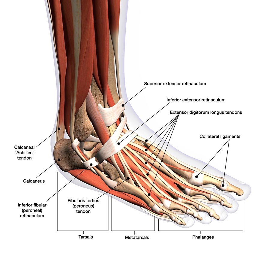
The foot is divided into three parts:
- The forefoot consists of five toes (phalanges) and five longer bones (metatarsals).
- The midfoot forms the arch and consists of the three cuneiform, cuboid and navicular bones.
- The hindfoot forms the heel and ankle. The calcaneus is the heel bone. The ankle is described below.
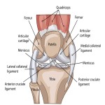
The ankle is formed of the talus bone, which supports the tibia (shin bone) and fibula in the leg. The tibiotalar joint (ankle joint) allows the foot to move up and down. The lateral malleolus is the bony protrusion on the outer ankle, formed by the distal end of the fibula. The medial malleolus is the inner ankle bone, formed by the distal end of the tibia.
Cartilage cushions the bones and allows them to glide smoothly over one another. Tendons connect muscles to bone to provide support. The Achilles tendon, which wraps around the heel bone, is the largest and strongest tendon in the body. Bursae, small sacs that contain synovial fluid, help to decrease friction between tendons and bones or skin.
Ligaments connect bones to other bones. The plantar fascia is the longest foot ligament, acting as a shock absorber and supporting the foot arch. Other ligaments include the talo-fibular and calcaneo-fibular ligaments.
There are 20 muscles in the foot, categorised as intrinsic (responsible for toe movement) and extrinsic (located in the lower leg and responsible for foot movement).