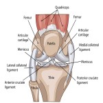


The knee joint is made up of two parts. The part of the knee between the end of the thigh bone (femur) and the top of the shin bone (tibia) is called the tibiofemoral joint. The patellofemoral joint is between the end of the thigh bone (femur) and the kneecap (patella).
The knee joint is surrounded by synovial fluid which keeps it lubricated. The bones are covered by smooth joint surface (articular) cartilage that allows them to glide smoothly together without friction. If the joint surface is damaged through wear and tear or a knee injury, arthritis can develop.
Cruciate ligaments
These are found inside your knee joint. They cross each other to form an “X” with the anterior cruciate ligament in front and the posterior cruciate ligament at the back. The cruciate ligaments control the back and forth motion of your knee.
Collateral ligaments
These are found on the sides of your knee. The medial or “inside” collateral ligament (MCL) connects the femur to the tibia. The lateral or “outside” collateral ligament (LCL) connects the femur to the smaller bone in the lower leg (fibula). The collateral ligaments control the sideways motion of your knee and brace it against unusual movement.
There are two meniscal cartilages in the knee that act as shock absorbers – one on the inner and one on the outer side. They sit between the curved lower part of the thigh bone and the flat upper part of the shin bone. Their job is to evenly distribute the load from the thigh bone to shin bone when walking and to provide knee stability. If the menisci are damaged, this can cause the cartilage beneath to become damaged and develop arthritis.